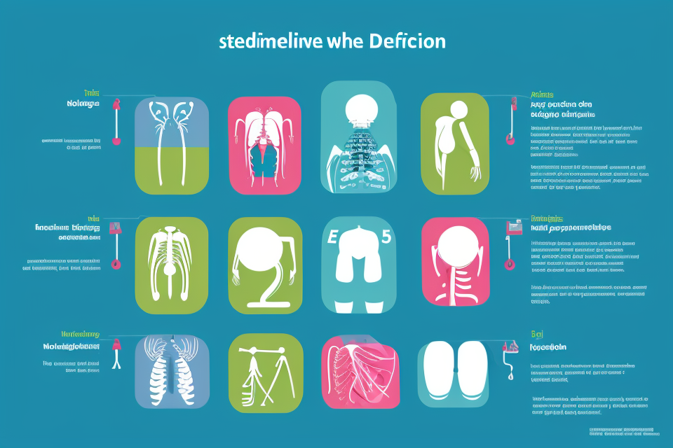
What are the 4 Stages of Spinal Degeneration?
Your spine is a vital part of your body that plays a crucial role in supporting your weight, protecting your spinal cord, and allowing for movement. However, as you age, your spine can degenerate, leading to various problems such as back pain, numbness, and weakness. Understanding the four stages of spinal degeneration can help you recognize the early signs and take proactive measures to prevent further damage. In this article, we will discuss the four stages of spinal degeneration and what you can do to maintain a healthy spine. So, let’s dive in!
The four stages of spinal degeneration are: 1) Degenerative disc disease: characterized by herniated or bulging discs, narrowing of the spinal canal, and nerve compression. 2) Spinal stenosis: narrowing of the spinal canal causing compression of the spinal cord and nerves. 3) Osteoarthritis: degeneration of the joints and bones in the spine leading to pain and stiffness. 4) Fusion: abnormal growths or bony overgrowths that can cause compression of the spinal cord and nerves. Each stage can vary in severity and symptoms, and treatment options will depend on the specific stage and underlying cause of the spinal degeneration.
Stage 1: Degenerative Disc Disease
Disc Anatomy and Function
The intervertebral discs are the small, spongy structures that are situated between the vertebrae of the spine. These discs serve a crucial purpose in the spine, as they act as shock absorbers and allow for flexibility and movement of the spine.
Each disc is composed of two main parts: the annulus fibrosus and the nucleus pulposus. The annulus fibrosus is the outer layer of the disc and is made up of layers of fibers that surround the nucleus pulposus, which is the inner, softer part of the disc.
The nucleus pulposus is responsible for the ability of the disc to absorb shock and provide flexibility to the spine. It is made up of a high water content and can change shape and size depending on the movement and position of the spine.
In summary, the intervertebral discs play a vital role in the function of the spine by acting as shock absorbers and allowing for flexibility and movement. Understanding the anatomy and function of these discs is important in understanding degenerative disc disease and the progression of spinal degeneration.
Causes of Degenerative Disc Disease
Aging is a major contributing factor to degenerative disc disease, as the discs in the spine naturally degenerate over time. As the discs lose their elasticity and hydration, they become less able to absorb shock and provide cushioning for the spinal vertebrae.
Genetics also play a role in the development of degenerative disc disease. Certain genetic factors can make individuals more susceptible to disc degeneration, as well as influence the rate at which it occurs.
Poor posture can also contribute to the development of degenerative disc disease. Prolonged periods of sitting or standing in a slouched position can place excessive stress on the discs in the spine, leading to damage and degeneration over time.
Trauma to the spine, such as from a car accident or fall, can also cause degenerative disc disease. These types of injuries can damage the discs and lead to changes in their structure and function, increasing the risk of further degeneration and damage over time.
Symptoms of Degenerative Disc Disease
Degenerative disc disease is a condition that affects the spine, specifically the intervertebral discs that cushion the vertebrae. As the discs degenerate, they can cause a range of symptoms, including pain, radiculopathy, weakness, and reduced range of motion.
Pain
Pain is a common symptom of degenerative disc disease. The pain can be described as dull, aching, or sharp, and it is usually felt in the back or neck. The pain may be constant or intermittent, and it can range from mild to severe.
Radiculopathy
Radiculopathy is a condition in which the nerves that exit the spine are compressed or irritated. This can cause pain, tingling, and numbness in the arms or legs. Radiculopathy is a common symptom of degenerative disc disease, and it can be particularly severe if the disc is pressing on a nerve root.
Weakness
Weakness can also be a symptom of degenerative disc disease. The weakness may be felt in the arms or legs, and it can be caused by the compression of nerves or by the degeneration of the discs themselves.
Reduced range of motion
Finally, degenerative disc disease can also cause a reduced range of motion in the back or neck. This can make it difficult to perform certain activities, such as bending or twisting, and it can also make it difficult to maintain good posture.
Overall, the symptoms of degenerative disc disease can have a significant impact on a person’s quality of life. If you are experiencing any of these symptoms, it is important to seek medical attention to determine the best course of treatment.
Diagnosis of Degenerative Disc Disease
Degenerative disc disease is a condition that affects the spine, specifically the intervertebral discs that act as shock absorbers between the vertebrae. Accurate diagnosis is crucial for effective treatment, and a combination of physical examination and imaging studies is typically used to confirm the presence of degenerative disc disease.
Physical examination plays a significant role in diagnosing degenerative disc disease. A doctor will perform a thorough evaluation of the patient’s back, assessing range of motion, reflexes, and sensation. They may also perform a neurological exam to determine if the nerve damage has occurred. Additionally, the doctor may ask the patient to perform certain movements or exercises to help identify any sources of pain or discomfort.
Imaging studies are also essential in diagnosing degenerative disc disease. X-rays are often used to evaluate the spine and determine if there are any changes in the bones or vertebrae. However, X-rays are not always reliable in detecting disc problems, as they only provide a limited view of the spine.
Magnetic resonance imaging (MRI) is a more advanced imaging technique that can provide detailed images of the spine and discs. An MRI can reveal herniated discs, disc degeneration, and other structural problems in the spine. Computed tomography (CT) scans may also be used in conjunction with MRIs to provide a more comprehensive view of the spine.
In conclusion, an accurate diagnosis of degenerative disc disease requires a combination of physical examination and imaging studies. A doctor will evaluate the patient’s symptoms, perform a physical examination, and order imaging studies to determine the extent of the damage and develop an appropriate treatment plan.
Treatment Options for Degenerative Disc Disease
Conservative Treatment
Conservative treatment options for degenerative disc disease are non-invasive and aim to manage pain and prevent further damage to the disc. These treatments include:
- Pain Medications: Over-the-counter pain relievers such as acetaminophen or nonsteroidal anti-inflammatory drugs (NSAIDs) may be recommended to manage pain. In some cases, prescription pain medications may be prescribed.
- Physical Therapy: A physical therapist may recommend exercises to strengthen the muscles around the affected disc, improve flexibility, and reduce pain.
- Heat and Cold Therapy: Applying heat or cold to the affected area can help alleviate pain and reduce inflammation.
- Massage Therapy: Massage therapy may help reduce muscle tension and improve circulation, which can help manage pain.
Minimally Invasive Procedures
Minimally invasive procedures are surgical interventions that use small incisions and minimal disruption of surrounding tissues. These procedures may be recommended if conservative treatments have not provided sufficient relief. Some minimally invasive procedures for degenerative disc disease include:
- Cervical or Lumbar Disc Replacement: In this procedure, a damaged disc is replaced with an artificial disc that helps restore natural disc function.
- Vertebroplasty or Kyphoplasty: These procedures involve injecting a bone-like material into the affected vertebrae to help stabilize the spine and reduce pain.
Surgical Intervention
In some cases, surgical intervention may be necessary to address degenerative disc disease. Surgical options include:
- Discectomy: In this procedure, a portion of the damaged disc is removed to relieve pressure on surrounding nerves.
- Spinal Fusion: This procedure involves joining two or more vertebrae together to stabilize the spine and reduce pain.
- Artificial Disc Replacement: Similar to cervical or lumbar disc replacement, this procedure involves replacing a damaged disc with an artificial disc to restore natural disc function.
It is important to note that the appropriate treatment option for degenerative disc disease will depend on the severity of the condition and the individual’s specific needs. Consulting with a healthcare professional is essential to determine the most appropriate treatment plan.
Stage 2: Spinal Stenosis
Definition and Causes of Spinal Stenosis
Description of Spinal Stenosis
Spinal stenosis is a condition in which the spinal canal narrows, putting pressure on the spinal cord and nerves. This narrowing can be caused by a variety of factors, including herniated discs, thickened ligaments, or the growth of bone spurs. As a result, the spinal cord and nerves can become compressed, leading to pain, numbness, and weakness in the affected area.
Causes of Spinal Stenosis
The narrowing of the spinal canal that causes spinal stenosis can be caused by a variety of factors, including:
- Herniated discs: The soft, jelly-like center of a spinal disc can bulge out of place and press against the spinal cord or nerves.
- Thickened ligaments: The ligaments that hold the spinal vertebrae together can become thick and stiff, narrowing the spinal canal.
- Bone spurs: Bony growths that develop on the vertebrae can also contribute to the narrowing of the spinal canal.
- Age-related wear and tear: Over time, the spinal discs and ligaments can degenerate, leading to the development of spinal stenosis.
- Trauma: A sudden injury to the spine, such as a car accident or fall, can also cause spinal stenosis.
Symptoms of Spinal Stenosis
Spinal stenosis is a condition that occurs when the spinal canal narrows, putting pressure on the spinal cord and nerves. This can cause a range of symptoms, including:
- Radiculopathy: Radiculopathy occurs when the nerves that exit the spine are compressed or irritated. This can cause pain, tingling, and weakness in the arms or legs.
- Weakness: Weakness can occur in the arms or legs as a result of nerve compression. This can make it difficult to perform daily activities that require the use of the affected limbs.
- Pain: Pain is a common symptom of spinal stenosis. The pain can be felt in the back, neck, or limbs, and may be described as aching, sharp, or burning.
- Numbness: Numbness can occur in the fingers, toes, or other parts of the body, and can be accompanied by tingling or a feeling of pins and needles.
It is important to note that not everyone with spinal stenosis will experience all of these symptoms. The severity of symptoms can also vary widely from person to person. If you are experiencing any of these symptoms, it is important to see a doctor for a proper diagnosis and treatment plan.
Diagnosis of Spinal Stenosis
Diagnosing spinal stenosis typically involves a combination of physical examination and imaging studies. During a physical examination, a healthcare provider will assess a patient’s range of motion, reflexes, and overall neurological function. This examination can help identify any abnormalities or areas of pain and can help determine if a patient’s symptoms are consistent with spinal stenosis.
Imaging studies are also an important part of the diagnosis of spinal stenosis. X-rays can provide a clear view of the bones in the spine and can help identify any changes or abnormalities. Magnetic resonance imaging (MRI) is another commonly used imaging study for spinal stenosis. An MRI can provide detailed images of the spinal cord and surrounding structures, allowing healthcare providers to see any changes or damage to the spinal cord. Computed tomography (CT) scans may also be used to provide detailed images of the spine and can help identify any other conditions that may be contributing to a patient’s symptoms.
In addition to these imaging studies, healthcare providers may also order blood tests to check for any underlying conditions or infections that may be contributing to a patient’s symptoms.
Overall, the diagnosis of spinal stenosis typically involves a thorough physical examination and a combination of imaging studies to identify any changes or abnormalities in the spine. By identifying spinal stenosis early, healthcare providers can work with patients to develop a treatment plan to manage their symptoms and prevent further damage to the spine.
Treatment Options for Spinal Stenosis
Conservative treatment options are typically the first course of action for patients with spinal stenosis. These treatments aim to relieve pain and other symptoms without resorting to invasive procedures or surgery. Conservative treatments may include:
- Pain Medication: Over-the-counter pain relievers such as ibuprofen or acetaminophen may help alleviate pain associated with spinal stenosis. In some cases, a doctor may prescribe stronger pain medication to manage more severe pain.
- Physical Therapy: A physical therapist can develop an exercise program tailored to the patient’s specific needs. This may include stretching exercises to improve flexibility, strengthening exercises to build muscle, and aerobic exercises to improve overall fitness.
- Activity Modification: A doctor may recommend modifying activities that exacerbate symptoms, such as avoiding prolonged standing or sitting. Alternating between sitting and standing may help alleviate pressure on the spine.
Minimally invasive procedures are generally considered when conservative treatments are not effective in managing symptoms. These procedures aim to relieve pressure on the spinal nerves and may include:
- Epidural Steroid Injections: Steroid injections can help reduce inflammation and swelling around the affected nerve roots. These injections are typically administered in a physician’s office and may provide temporary relief for up to several weeks or months.
- Lumbar Epidural Nerve Blocks: Similar to epidural steroid injections, lumbar epidural nerve blocks involve the injection of a local anesthetic and steroid medication into the epidural space around the affected nerve root. This can help alleviate pain and discomfort associated with spinal stenosis.
In some cases, surgical intervention may be necessary to address spinal stenosis. Surgical options may include:
- Laminectomy: A laminectomy involves removing a portion of the vertebra (lamina) that is causing pressure on the spinal nerves. This procedure can provide significant relief from symptoms, but may also result in instability of the spine and require additional surgery to address.
- Discectomy: A discectomy involves removing a portion of a herniated disc that is pressing on a nerve root. This procedure can provide relief from leg pain and discomfort associated with spinal stenosis.
- Spinal Fusion: In some cases, a spinal fusion may be necessary to stabilize the spine after a laminectomy or discectomy. This procedure involves fusing two or more vertebrae together using bone graft material, metal hardware, or both.
It is important to note that surgery should only be considered after conservative treatments and minimally invasive procedures have been attempted and have not provided adequate relief. The decision to undergo surgery should be made in consultation with a qualified spine specialist.
Stage 3: Herniated Disk
Definition and Causes of Herniated Disk
A herniated disk occurs when the soft, gel-like center of a spinal disk bulges out through a crack or tear in the tough, outer layer of the disk. This can put pressure on the surrounding nerves, which can cause pain, numbness, and weakness in the affected area.
There are several factors that can contribute to the development of a herniated disk, including:
- Age: As we age, the disks in our spine naturally degenerate and lose some of their elasticity, making them more prone to herniation.
- Genetics: Some people may be more genetically predisposed to developing herniated disks.
- Physical activity: Engaging in activities that put repetitive stress on the spine, such as heavy lifting or long periods of sitting, can increase the risk of herniated disks.
- Trauma: A sudden impact or injury to the spine can also cause a herniated disk.
Understanding the causes of herniated disks can help individuals take steps to prevent them, such as maintaining good posture, avoiding heavy lifting, and engaging in regular exercise to keep the spine strong and flexible.
Symptoms of Herniated Disk
- Pain:
- In the lower back or neck
- May radiate to the legs or arms
- May be constant or intermittent
- Radiculopathy:
- Pain, tingling, or numbness that radiates along the nerve pathway
- Typically affects the extremities, such as the hands or feet
- May be accompanied by weakness or loss of sensation
- Weakness:
- Loss of strength or muscle power
- May affect the legs, arms, or shoulders
- May make it difficult to perform daily activities
- Numbness:
- Loss of sensation or feeling
- May affect the legs, feet, or hands
- May be accompanied by tingling or prickling sensations
In summary, the symptoms of a herniated disk in the spine can include pain, radiculopathy, weakness, and numbness. These symptoms may vary in severity and location, and may affect a person’s ability to perform daily activities. If you are experiencing any of these symptoms, it is important to seek medical attention to determine the cause and appropriate treatment.
Diagnosis of Herniated Disk
When diagnosing a herniated disk, a healthcare professional will typically begin with a physical examination. During this examination, the healthcare professional will ask questions about the patient’s symptoms and perform a series of tests to assess the patient’s range of motion, reflexes, and sensation. This helps the healthcare professional determine which nerves may be affected by the herniated disk.
In addition to a physical examination, imaging studies are often used to confirm the diagnosis of a herniated disk. There are several types of imaging studies that can be used, including X-rays, MRI, and CT scans.
X-rays are a type of imaging study that uses radiation to create images of the body’s internal structures. X-rays can help healthcare professionals identify bone spurs or other changes in the bones of the spine that may be causing symptoms.
MRI, or magnetic resonance imaging, is a type of imaging study that uses a strong magnetic field and radio waves to create detailed images of the body’s internal structures. MRIs are particularly useful for identifying herniated disks and other soft tissue injuries.
CT scans, or computed tomography scans, are another type of imaging study that uses radiation to create images of the body’s internal structures. CT scans are particularly useful for identifying fractures or other bone injuries.
In addition to these imaging studies, healthcare professionals may also order electromyography (EMG) to diagnose a herniated disk. EMG is a test that measures the electrical activity of muscles. This test can help healthcare professionals determine which nerves are affected by the herniated disk and can help guide treatment decisions.
Treatment Options for Herniated Disk
Conservative treatment options are typically the first course of action for patients with a herniated disk. These treatments aim to alleviate pain and inflammation without the need for invasive procedures. Conservative treatments may include:
- Rest: Limiting physical activity and avoiding positions that exacerbate pain may help reduce inflammation and promote healing.
- Ice and heat therapy: Applying ice to the affected area for 15-20 minutes at a time, several times a day, can help reduce pain and inflammation. Heat therapy may also be used to promote blood flow and reduce stiffness.
- Over-the-counter pain medications: Nonsteroidal anti-inflammatory drugs (NSAIDs) such as ibuprofen or naproxen can help reduce pain and inflammation.
- Physical therapy: A physical therapist can provide exercises and stretches to help improve spinal mobility and reduce pain.
- Massage therapy: Massage therapy may help reduce muscle tension and promote relaxation, which can help alleviate pain.
If conservative treatments do not provide sufficient relief, minimally invasive procedures may be considered. These procedures typically involve the use of needles or small incisions to access the affected area, rather than large incisions that can damage surrounding tissues. Examples of minimally invasive procedures for herniated disks include:
- Epidural steroid injections: Corticosteroids are injected into the epidural space around the affected nerve root to reduce inflammation and promote healing.
- Facet joint injections: Corticosteroids are injected into the facet joints to reduce inflammation and pain.
- Nerve root blocks: Corticosteroids are injected around the affected nerve root to reduce inflammation and pain.
In some cases, surgical intervention may be necessary to treat a herniated disk. Surgery may be recommended if conservative treatments have not provided sufficient relief, or if the herniated disk is causing significant nerve damage. Examples of surgical procedures for herniated disks include:
- Discectomy: The surgeon removes a portion of the herniated disk that is pressing on the affected nerve root.
- Laminectomy: The surgeon removes a portion of the vertebra to relieve pressure on the affected nerve root.
- Spinal fusion: The surgeon fuses two or more vertebrae together to stabilize the spine and reduce pressure on the affected nerve root.
It is important to note that surgery carries risks and should only be considered if other treatment options have been exhausted and the benefits of surgery outweigh the risks.
Stage 4: Spinal Fusion
Definition and Causes of Spinal Fusion
Spinal fusion is a surgical procedure that involves joining two or more vertebrae in the spine, thereby eliminating the movement between them. This is done to stabilize the spine and reduce pain caused by spinal degeneration.
Causes of spinal fusion:
- Spinal degeneration: As the spine ages, the discs between the vertebrae lose their elasticity, causing the space between the vertebrae to narrow. This can result in pressure on the nerves, leading to pain and discomfort.
- Trauma: A traumatic injury to the spine, such as a car accident or a fall, can cause fractures or dislocations that require spinal fusion to stabilize the spine.
- Inflammatory diseases: Conditions such as ankylosing spondylitis or rheumatoid arthritis can cause inflammation in the spine, leading to pain and stiffness. In some cases, spinal fusion may be necessary to relieve these symptoms.
- Congenital abnormalities: Some people are born with abnormalities in the spine, such as spina bifida or congenital curvature of the spine, which can require spinal fusion to correct.
In summary, spinal fusion is a surgical procedure that involves joining two or more vertebrae in the spine to stabilize the spine and reduce pain caused by spinal degeneration, trauma, inflammatory diseases, or congenital abnormalities.
Symptoms of Spinal Fusion
- Pain: Individuals experiencing spinal fusion may experience pain in the affected area. This pain can range from mild discomfort to severe pain that can interfere with daily activities.
- Reduced range of motion: As the spine undergoes fusion, the affected vertebrae become rigid and do not move as freely as they did before. This can lead to a reduced range of motion in the affected area.
- Weakness: Weakness can occur in the muscles surrounding the affected area, as the spinal fusion can affect the nerves that control these muscles. This weakness can make it difficult to perform physical activities and can interfere with daily living.
- Instability: The spine is designed to provide stability to the body, but spinal fusion can cause instability in the affected area. This can lead to an increased risk of falls and other injuries.
Diagnosis of Spinal Fusion
The diagnosis of spinal fusion involves a physical examination and imaging studies to assess the condition of the spine. The physical examination includes a thorough evaluation of the patient’s medical history, symptoms, and a series of tests to assess range of motion, strength, and reflexes.
Imaging studies are also performed to evaluate the spine and identify any abnormalities. X-rays are typically the first imaging study ordered, as they can provide a detailed view of the bones and joints in the spine. However, X-rays may not always reveal the extent of spinal degeneration, and other imaging studies such as MRI or CT scan may be necessary to provide a more comprehensive view of the spine.
The information gathered from the physical examination and imaging studies is used by the healthcare provider to determine the appropriate course of treatment for the patient. In some cases, spinal fusion surgery may be recommended to alleviate symptoms and improve spinal function. However, the decision to undergo surgery should be made on an individual basis, taking into account the patient’s overall health and the specifics of their condition.
Treatment Options for Spinal Fusion
Spinal fusion is the fourth stage of spinal degeneration, which occurs when the discs between the vertebrae in the spine have worn down, and the space between the vertebrae has narrowed. This stage is characterized by chronic pain, stiffness, and limited mobility. Treatment options for spinal fusion depend on the severity of the condition and the underlying cause.
Conservative treatment options are the first line of treatment for spinal fusion. These options include pain medication, physical therapy, and exercise. Pain medication can help relieve pain and reduce inflammation. Physical therapy can help improve flexibility and strengthen the muscles around the affected area. Exercise can also help improve range of motion and reduce pain.
Minimally invasive procedures are another option for treating spinal fusion. These procedures involve the use of small incisions and specialized instruments to access the affected area. Examples of minimally invasive procedures include microdiscectomy, laminectomy, and foraminotomy. These procedures can help relieve pressure on the nerves and alleviate pain.
Surgical intervention is usually reserved for cases where conservative and minimally invasive treatments have not been effective. Surgical options for spinal fusion include spinal fusion surgery, laminectomy, and discectomy. Spinal fusion surgery involves fusing the affected vertebrae together using bone grafts or metal implants. Laminectomy involves removing a portion of the vertebrae to relieve pressure on the nerves. Discectomy involves removing a portion of a herniated disc to relieve pressure on the nerves.
Overall, the choice of treatment option depends on the severity of the condition and the underlying cause. Conservative and minimally invasive treatments are typically preferred over surgical intervention, as they are less invasive and have fewer risks and complications. However, surgical intervention may be necessary in some cases to relieve severe pain and improve mobility.
FAQs
1. What are the four stages of spinal degeneration?
The four stages of spinal degeneration are:
1. Degenerative disc disease: This is the earliest stage of spinal degeneration. It occurs when the intervertebral discs, which act as shock absorbers between the vertebrae, begin to wear down. This can cause pain and stiffness in the back and neck.
2. Spinal stenosis: As the degeneration progresses, the space around the spinal cord begins to narrow. This can put pressure on the spinal cord and nerves, causing pain, weakness, and numbness in the legs and arms.
3. Facet joint arthritis: The facet joints, which connect the vertebrae, can become inflamed and arthritic. This can cause pain in the back and neck, and may limit mobility.
4. Spondylolisthesis: In this advanced stage of spinal degeneration, a vertebra can slip out of place, which can cause pain and may require surgery.
2. What are the symptoms of spinal degeneration?
The symptoms of spinal degeneration can vary depending on the stage of the condition. In the early stages, people may experience back pain and stiffness. As the condition progresses, people may experience pain, weakness, and numbness in the legs and arms, as well as difficulty walking or standing for long periods. In the advanced stages, people may experience loss of bowel or bladder control.
3. What causes spinal degeneration?
Spinal degeneration is caused by the natural wear and tear that occurs over time. As people age, the discs between the vertebrae can shrink and lose elasticity, which can cause pain and stiffness. Other factors that can contribute to spinal degeneration include genetics, obesity, and a sedentary lifestyle.
4. How is spinal degeneration treated?
Treatment for spinal degeneration depends on the stage of the condition and the severity of the symptoms. In the early stages, conservative treatments such as physical therapy, pain medication, and chiropractic care may be effective. In more advanced stages, surgery may be necessary to relieve pressure on the spinal cord and nerves. It is important to consult with a healthcare professional to determine the best course of treatment for your individual needs.